
Modeling the contribution of harmonic lattice vibrations to the diffuse signal with GOODVIBES
When a protein crystal is bombarded with X-rays, the resulting signal on the detector contains a wealth of information that is connected to the structure and dynamics of the crystal. The clearest structural information is contained within the bright, regularly-spaced Bragg peaks. These contain information about the average structure; the space group to which the crystal belongs (related to how proteins have packed into the unit cell) and the average positions of each atom can be determined from these intense spots. The diffuse signal, on the other hand, comes from all deviations of atoms from their average positions. Conformational heterogeneity1, crystal defects, and thermal motion all contribute to the diffuse signal and lead to their own characteristic features. The inverse problem of extracting information from this diffuse signal is one of the overarching goals of the DiffUSE collaboration.
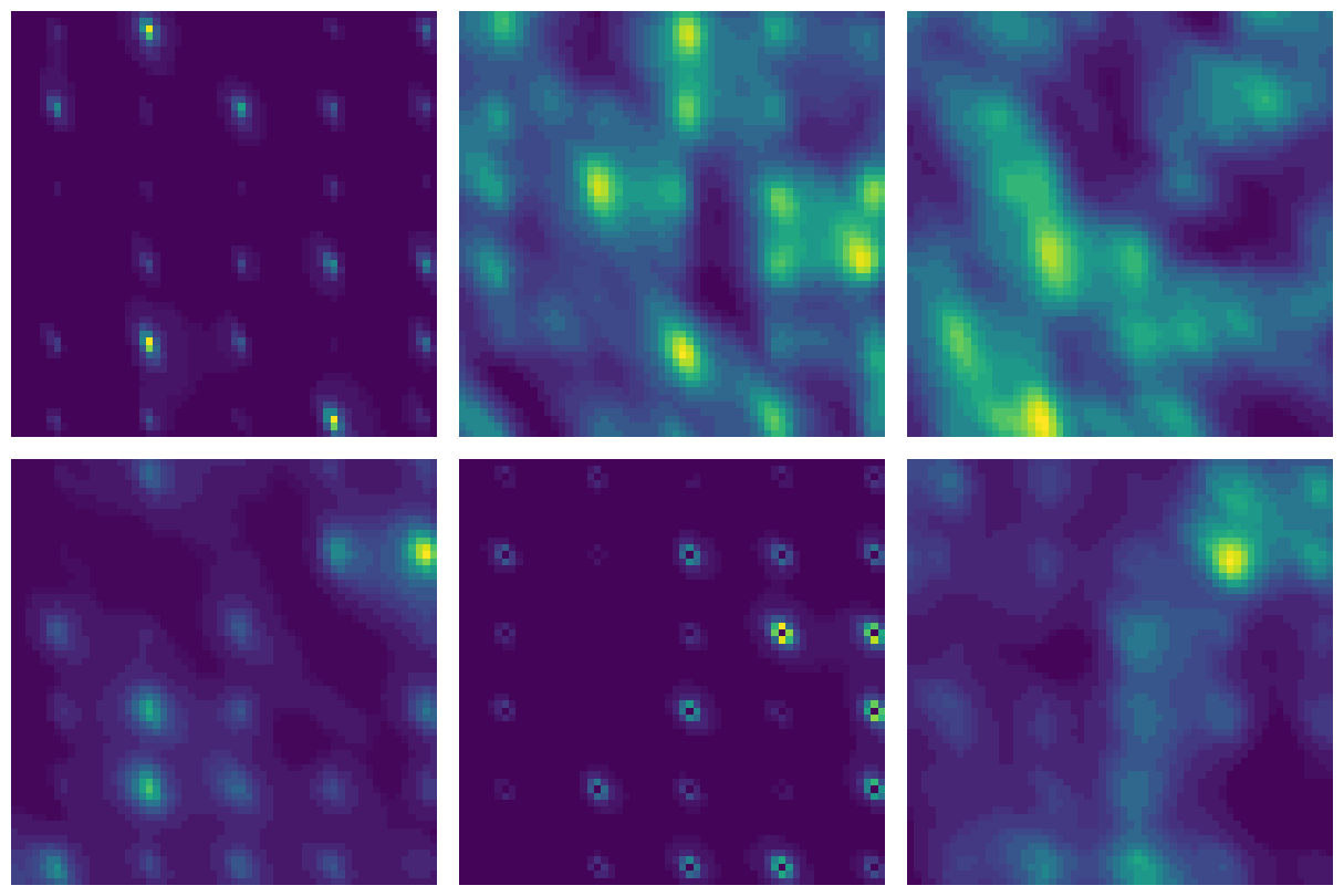
Figure 1: A moving slice through reciprocal space of the diffuse signal predicted by a model that includes lattice vibrations of a triclinic lysozyme crystal. The scale is different in each frame so that the diffuse signal between the Bragg peaks is visible.
The brightest features in the diffuse signal occur around the Bragg peaks, and are termed “halos” for their round (but often asymmetric) shape. These halos are well-reproduced by a model that includes thermally excited lattice vibrations, or phonons. Phonons are elastic waves that propagate through crystals and displace proteins from their average positions. Very low-frequency and long-wavelength phonons contain information about the often anisotropic sound speeds and elastic moduli of the crystal, but they do not contain direct information about internal protein motions. High-frequency and short-wavelength phonons can involve correlated motions of proteins in the crystal that are related to the biological function of isolated proteins in vivo.
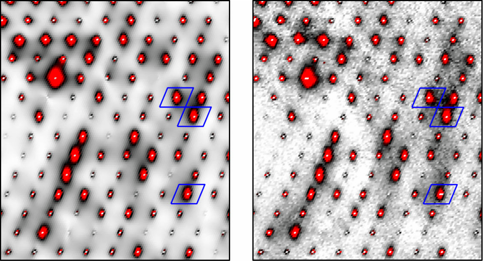
Figure 2: Selected regions of the diffuse X-ray scattering of a lattice dynamics model (left) and of a real protein crystal (right). The red features are halos. The halos which are surrounded by blue boxes were used to refine the model to the data (image from Meisburger et al. 20202).
At any temperature, the most populated phonon modes are the lowest-frequency ones. Unsurprisingly, then, these low-frequency phonons which only contain information about long-wavelength crystal properties produce the halos and dominate the diffuse signal3. General Optimization Of Diffuse halos from VIBrational Elastic network Simulations (GOODVIBES) is a software package that produces a full forward model of the phononic contribution to the diffuse signal4. It works by modeling a protein crystal as a periodic collection of rigid bodies which are connected to neighbors by springs. From this model and an independently refined map of the electron density, information about the expected diffuse signal contributed by thermally excited phonons can be readily computed. The strengths of springs that connect adjacent rigid bodies are adjusted until the diffuse signal from the model well-fits the bright, halo-like features of the experimental data.
As a full forward model of the phononic contribution to diffuse scattering, the utility of GOODVIBES is many-fold and leads to several open questions:
- If we model the long-wavelength contribution to the diffuse signal very well, can we subtract it out to extract biologically relevant motions that contribute to the diffuse signal?
- After the halos are fit, can we understand the more complicated, structured diffuse signal between the Bragg peaks as also coming partially from phonons?
- Can more sophisticated modeling of the internal motions of the protein be developed so that harmonic modes related to biological function can be fit to the experimental data?
Where we are now
We are re-developing the GOODVIBES pipeline using entirely open-source Python packages. As part of this, we are re-thinking each step in the pipeline, from the structure refinement to the parameterization of the interactions within the asymmetric unit of the crystal. We want the refinement procedure to be flexible enough to handle protein crystals in any space group and of any unit cell size. While we are aiming to refine an elastic network model of Mac1 in the P43 space group (PDB 7tx05) to experimental diffuse scattering data, here we show results based on the refinement of lysozyme in the P1 space group (PDB 6o2h6) from an older iteration of GOODVIBES as proof of concept. We treat the entire protein in the unit cell as a rigid body, so no internal protein modes are modeled. With one protein per unit cell, there are consequently 6 phonon bands, which change shape appreciably during the refinement process.
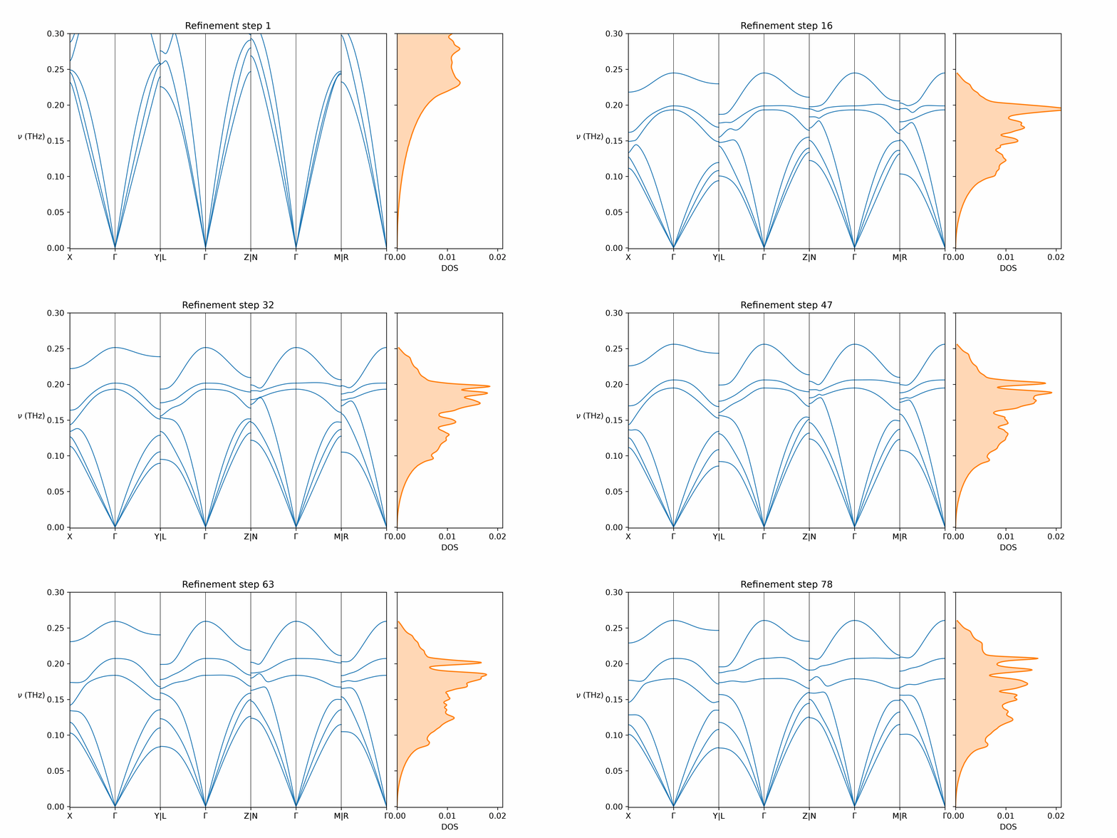
Figure 3: The phonon band structure (left) and density of states (right) of triclinic lysozyme predicted by GOODVIBES during refinement. Each changes dramatically during refinement to fit the halos in the experimental data.
After refinement, the halos are well-fit, and the fully refined harmonic model contains a wealth of easily extractable information that can be compared with other experimental data: the phonon band structure and density of states allow us to determine the independent elastic moduli of the crystal and to calculate the phononic contribution to thermodynamic quantities like entropy and specific heat, which may be anomalous7.
Where we’re going next
We have a combination of short- and long-term goals for the future development of GOODVIBES.
- Demonstrate the full refinement pipeline for the Mac1 protein crystal in the P43 space group (PDB 7tx05), modeling the two independent chains in the asymmetric unit as separate rigid bodies.
- Incorporate structure refinement into the pipeline - can information contained within the diffuse signal be used to get a better average structure?
- Rethink the parameterization of the rigid body interactions - are springs between residues found with a neighbor search an optimal choice for this refinement problem?
- Incorporate flexibility in the rigid body decomposition, perhaps integrating it with other TLS refinement software. This will allow further exploration of how model parameterization affects the portions of the diffuse signal not included in the fit to the experimental diffuse signal.
As we continue to develop GOODVIBES, we will be thinking more deeply about how to integrate information gleaned from a fit to this model which assumes phonons in regular crystals generate the diffuse signal with other models that include contributions that come from motion that cannot be understood as harmonic. Non-harmonic contributions to the signal include conformational heterogeneity (modeled with sampleworks8) and highly disordered protein and solvent (found through molecular dynamics simulations (e.g. Wall et al. 20199)).
References:
-
Wankowicz, S. A. (2026). Recovering Conformational Heterogeneity from the Protein Data Bank at Scale. https://doi.org/10.82153/pff0-ck46 ↩
-
Meisburger, S.P., Case, D.A. & Ando, N. Diffuse X-ray scattering from correlated motions in a protein crystal. Nat Commun 11, 1271 (2020). https://doi.org/10.1038/s41467-020-14933-6 ↩
-
Polikanov YS, Moore PB. Acoustic vibrations contribute to the diffuse scatter produced by ribosome crystals. Acta Crystallogr D Biol Crystallogr. 2015 Oct;71(Pt 10):2021-31. doi: https://doi.org/10.1107/S1399004715013838 ↩
-
Meisburger, S. P., Case, D. A., & Ando, N. (2023). Robust total X-ray scattering workflow to study correlated motion of proteins in crystals. Nat Commun, 14(1), 1228. https://doi.org/10.1038/s41467-023-36734-3 ↩
-
Correy, G.J., Fraser, J.S. (2022). Crystal structure of SARS-CoV-2 NSP3 macrodomain in complex with ADP-ribose at pH 9 (P43 crystal form). doi: https://doi.org/10.2210/pdb7TX0/pdb ↩ ↩2
-
Meisburger, S.P., Ando, N. (2020). Hen lysozyme in triclinic space group at ambient temperature - diffuse scattering dataset. doi: https://doi.org/10.2210/pdb6O2H/pdb ↩
-
Meinhold, L., Merzel, F., & Smith, J. C. (2007) Lattice Dynamics of a Protein Crystal. Phys. Rev. Lett. 99, 138101. https://doi.org/10.1103/PhysRevLett.99.138101 ↩
-
Chrispens, K., Collins, M. D., Mai, D., Wankowicz, S. A., Fraser, J. S., & van den Bedem, H. (2026). sampleworks: A Modular Platform for Experimentally Guided Biomolecular Ensemble Generation. https://doi.org/10.82153/jkxj-tw08 ↩
-
Wall ME, Calabró G, Bayly CI, Mobley DL, Warren GL. Biomolecular Solvation Structure Revealed by Molecular Dynamics Simulations. J Am Chem Soc. 2019 Mar 20;141(11):4711-4720. https://doi.org/10.1021/jacs.8b13613 ↩
Comments